News & Media
Gold-coated iron oxide nanoparticles are a promising agent for cancer therapy and imaging
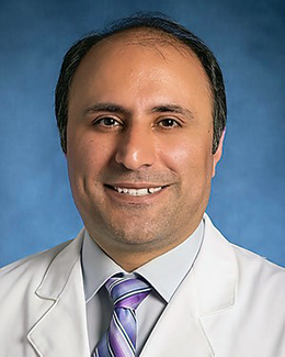
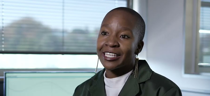
Professor Ali Shakeri-Zadeh
The UNESCO-UNISA Africa Chair in Nanoscience and Nanotechnology (U2ACN2), Professor Malik Maaza, recently hosted an informative webinar series. Ali Shakeri-Zadeh, an Assistant Professor of Radiology at Johns Hopkins University, presented his work. He discovered that gold-coated iron oxide nanoparticles are emerging as multi-functional nanomaterials for cancer nano theranostics applications.
The current and conventional methods for cancer treatment are surgery, radiation therapy, and chemotherapy. Other treatments include immunotherapy, hormone therapy, targeted therapy, and stem cell therapy. Surgery is an invasive method, while chemotherapy and radiation have severe side effects because they are not selective with cells. Therefore, they can cause damage to collateral normal cells. Nanoparticles can be an excellent solution to address the non-selectivity of cells. Theranostic nanoparticles are used for imaging and therapy. They can address both therapeutic applications and diagnostic applications.
Superparamagnetic (SPIO) nanoparticles have an application in Magnetic Resonance Imaging (MRI). SPIO nanoparticles can be used for the weighting of the Magnetic Resonance Images. There is an option to make different nanoparticles through SPIO nanoparticles. SPIO nanoparticles are also known for possessing superior magnetic properties with the capability of being functionalised with other ligands. In the field of cancer imaging and cancer therapy, radiotherapy can be performed to target gold nanoparticles (AuNP) in cancer cells through radiation. Gold nanoparticles can be used to target chemotherapy to cancer cells, and they can be used in immune and photothermal therapy.
Shakeri-Zadeh focused on gold-coated Ion oxide nanoparticles (NP) as theranostic nanoparticles. The combination of both can address different challenges that are encountered with image-guided therapy. The nanoparticles can be used for imaging such as Magnetic Resonance Imaging (MRI), Magnetic Particle Imaging (MPI), Photoacoustic Imaging (PAI), Magnetic-Photo-Acoustic (MPA) Imaging, and Computed Tomography (CT) scans. For therapy, there are options for Radiotherapy (RT), Photothermal therapy (PTT) and Magnetic Floyd Hyperthermia. In addition, dosimetry, distribution study and monitoring of the therapeutic and therapy sessions can occur.
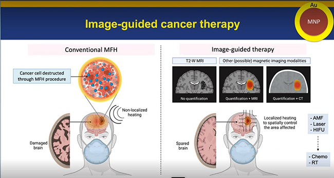
Magnetic Particle Imaging (MPI)-guided hyperthermia has a side effect in that there is a high chance of irradiating the regular part of the brain, so it can be damaged. However, suppose a Magnetic Resonance Imaging (MRI) or Computed Tomography (CT)-guided hyperthermia can occur. In that case, the therapy field can be localised to the region of interest and selectively destroy the brain tumour.
One promising solution for non-invasive imaging is image-based therapy using multi-functional nanoparticles such as gold-coated Ion oxide nanoparticles. It is a good agent for therapy and imaging.
* By Hanli Wolhuter, Communication and Marketing Specialist and Musa Buthelezi, Intern, College of Graduate Studies
Publish date: 2022-11-24 00:00:00.0

 Unisa's student leadership engage with Russian ambassador
 Re-igniting and re-imagining Pan Africanism, Afrocentricity and Afrofuturism in the 21st century
 Young Unisa science stars join elite Lindau Nobel Laureate group
 Education MEC addresses Unisa autism seminar
 Seven Unisans nominated for the NSTF-South32 Awards 2023/2024